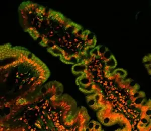
Channel drawing figure by Jalili-Firoozinezhad and al. (2019); Image adapted from S. Schuller (wellcome collection CC BY 4.0).
GUT-ON-A-CHIP PACK
All microfluidic pieces included, quick and easy assembly
Controlled shear stress under laminar flow, easy medium distribution
Improved incubation parameters closer to physiological conditions
Microfluidic gut-on-a-chip
Gut cell culture on a membrane in a microfluidic channel is now accessible using this gut-on-a-chip Pack. Seeded on a porous membrane, the cells are continuously perfused, mimicking the physiological gut environment for an improved cell culture that allows experiments in environments closer to in vivo than traditional 2D cell cultures.
Based on the high accuracy OB1 flow controller (Elveflow) and a porous membrane implemented inside a microfluidic chip, this solution contains all the required microfluidic pieces for researchers to set up the coculture of intestinal endothelial and epithelial cells. We will offer you support during your experiment setup if you are not familiar with microfluidic technologies.
Organ-on-chip experiments are difficult and complex to implement. We have collaborated on several organ-on-a-chip projects and accumulated valuable experience. We are always happy to exchange with researchers!
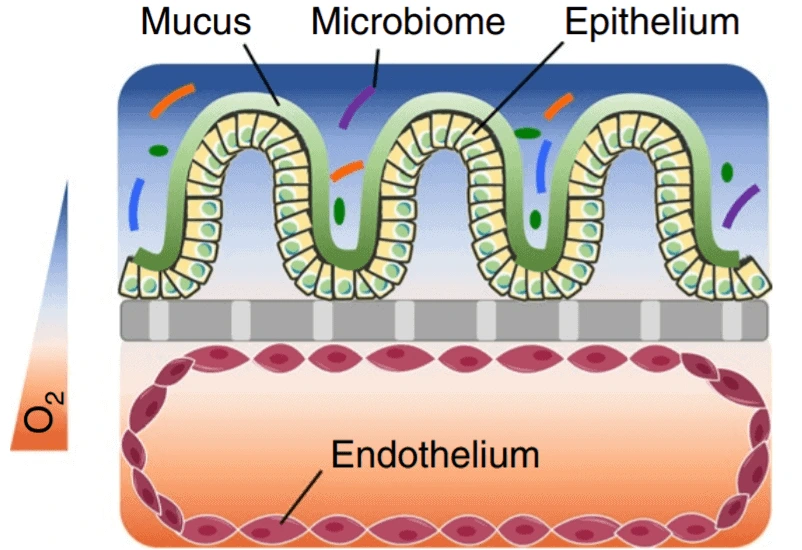
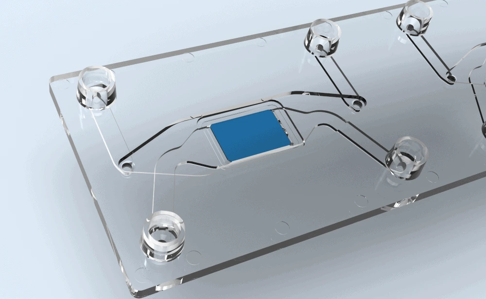
A standard microfluidic gut-on-a-chip culture pack contains a pumping channel coupled with a distributor to seed the cells on both sides of the membrane in the chip, creating an endothelial and an epithelial cell layer that can be used for new therapeutic developments or toxicity screening.
For example, the upper channel can be seeded with human umbilical cord vein endothelial cells (HUVECs) to form the endothelial cell layer, and Caco-2 cells (epithelial colorectal cells) can be planted at the lower perfusion chamber to form the epithelial layer. This intestine 3D cell structure, coupled with dynamic perfusion conditions, allows a physiologically relevant model to be created.
This Pack can also contain other instruments that can be adapted and modified depending on your specifications. It can also be applied to another commercially available or homemade microfluidic chip.
The MUX distributor is helpful for quickly injecting different substances like drugs, and a 3/2 valve is used to choose which channel to perfuse. The efficiency of the perfusion will be directly linked to the flow rate inside the upper and lower channels. Flow rates can be measured with the MFS or BFS flow rate sensors (Elveflow).
Gut-on-a-chip pack setup
This pack reproduces physiologically realistic shear stress variations, pressure, and strain. Read more about the subject in our gut-on-a-chip review.
An integrated pack assures good compatibility between the different components, allows you to start your experiment right off the shelf, is controlled by one software, and can be used for purposes other than gut-on-a-chip.
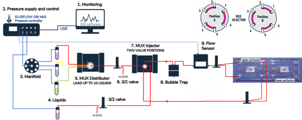
The gut-on-a-chip pack includes:
- An OB1 flow controller (Elveflow)
- A rotative distribution valve (MUX distributor, Elveflow)
- The MUX recirculation allows a close recirculation of the medium
- A microfluidic flow sensor (MFS or BFS, Elveflow)
- A bubble trap
- Cross-flow membrane chip from Microfluidic ChipShop or your homemade chip
- Falcon reservoirs
- 3 ports / 2 positions microfluidic valves
- A MUX wire (Elveflow) that allows the control of the valves
- A manifold pressure splitter
- Connectors, tubing, filters,…
- Software and SDK libraries (C++, Python, MATLAB, LabVIEW) (Elveflow)
- (HUVEC and Caco-2 cells if necessary)
Microfluidic gut-on-a-chip principle
Various research groups showed the importance of fluid flow to apply shear stress that mimics the conditions closest to the physiological one to grow the cells into villi structures [1-2]. Caco-2 epithelial cell growth into crypt and villus-like structures is significantly better in a microfluidic chip with a membrane than in a more classical transwell cell culture [3].
The crypt and villus-like structure cell layer morphology exhibits better activity in a microenvironment closer to a human gut and can support the growth of human intestine microbial flora [2, 4]. In a gut-on-a-chip, the dynamic culture enhances the extracellular matrix (ECM), which influences the surrounding microenvironment, creating an even more physiologically relevant gut epithelium cell layer model [5].
To summarize, the intestine on the chip system is a valuable model for physiology studies, drug development, and personalized medicine without using animal models [6].
Advantages of microfluidics for gut-on-a-chip
Microfluidics is an excellent way to decrease the potentially valuable samples used during an experiment, like a patient’s biopsy.
The combination of the OB1 flow controller, the MUX distributor, and the 3/2 valve coupled with the automation software allows the creation of very efficient and controlled experiments. The instruments contained in this pack allow optimal flow control, reactivity, and precision for the gut-on-a-chip cell culture experiment. At this scale, the different properties of the fluid can be precisely tuned in real time to stay close to the physiological environment.
Human organs on chips are more effective than 2D classical or animal models. The European Union and the public are pushing to reduce the use of animal models. Furthermore, organ chip development can lead to Personalized medicine by using stem cells directly from the patient.
Drug toxicity or pathogen effects on the intestine can be assessed using microfluidic gut-on-a-chip cell culture with more flexible, precise, and efficient experiments.
References
- Chi, Meiying, et al. “A microfluidic cell culture device (μFCCD) to culture epithelial cells with physiological and morphological properties that mimic those of the human intestine.” Biomedical microdevices 17 (2015): 1-10.
- Kim, Hyun Jung, and Donald E. Ingber. “Gut-on-a-Chip microenvironment induces human intestinal cells to undergo villus differentiation.” Integrative Biology 5.9 (2013): 1130-1140.
- Maurer, Michelle, et al. “A three-dimensional immunocompetent intestine-on-chip model as in vitro platform for functional and microbial interaction studies.” Biomaterials 220 (2019): 119396.
- Kim, Hyun Jung, et al. “Human gut-on-a-chip inhabited by microbial flora that experiences intestinal peristalsis-like motions and flow.” Lab on a Chip 12.12 (2012): 2165-2174.
- De Gregorio, Vincenza, et al. “Intestine‐on‐chip device increases ECM remodeling inducing faster epithelial cell differentiation.” Biotechnology and Bioengineering 117.2 (2020): 556-566.
- Bein, Amir, et al. “Microfluidic organ-on-a-chip models of human intestine.” Cellular and molecular gastroenterology and hepatology 5.4 (2018): 659-668.
- Jalili-Firoozinezhad, Sasan, et al. “A complex human gut microbiome cultured in an anaerobic intestine-on-a-chip.” Nature biomedical engineering 3.7 (2019): 520-531.
Customize your pack
Contact our experts if you have doubts about the setting that could perfectly fit your specific application!
The pack contains cross-flow membrane chips in polystyrene (PS) or cycloolefin copolymer (COP) materials. Two pore sizes are available: 8 µm and 0.2 µm. The channel can also be hydrophilized. Custom-specific membranes can also be made on demand.
The provided gut-on-a-chip pack can be highly personalized, and changes can be made on demand, such as replacing the MUX recirculation with cheaper microfluidic valves to perform the medium recirculation in the microfluidic channel but with increased complexity.
The OB1 MK4 flow controller can also have additional channels with different pressure ranges.
Coriolis flow sensors can replace the MFS flow sensors in the setup to obtain the best possible flow control.
Bubbles are highly detrimental to cell culture and should be avoided; a bubble remover can be added to the pack to prevent the cells from being exposed to air bubbles.
We assembled other packs for organ-on-chip, the blood-brain-barrier-on-a-chip, and the endothelial cell culture packs.
Contact our scientists for any questions about this gut-on-a-chip culture pack and how it can match your specifications.
– Check our other Packs for various applications –
Can I order a pack?
Since Packs are products that are still being developed, we have a few eligibility criteria to maximize their success rate. A discussion with our experts is needed to determine your specific needs to offer you a personalized response.
Is the setup sterilizable?
Yes, we have developed a simple protocol for sterilization and cleaning that is provided along with the user guide.
Can a pack be customized based on my specific application?
Yes! Our experts will establish which instruments are best suited for your application, such as the type of flow sensor or the number of flow controller channels you need to perform your experiment. Drop us a line at innovation@microfluidic.fr.
Can I buy individual instruments?
You can order the instruments on the product section of our website.