Search
Close this search box.

Drug testing: organ-on-chip models vs. standard models

Introduction to organ-on-chip models

In vitro and in vivo models are widely used to investigate human pathophysiology and toxicology studies. The goal of this review is to highlight the main advantages and limitations of each model and to show how the use of organ-on-chip model technology can address their drawbacks.

mouse organ-on-chip in-vitro in-vivo drug testing

The article refers specifically to lung systems (i.e., capillary-alveolar interface). Still, it also applies to other organs since the characteristics of joint in vivo/ in vitro systems result in being related to the system itself rather than the organ/ tissue studied.


You can look here for a description of the use of the alveolar-capillary barrier to design microfluidic lung-on-a-chip systems. To start using microfluidics to build lung-on-a-chip models, check out our pack.


You can continue reading here to learn more about the origin and design of lung-on-a-chip systems.

Overview of organ-on-chip in vitro and in vivo models

Organ-on-chip models vs. ANIMAL MODELS [1, 2]

mouse-organ-on-chip-in vitro-in vivo-drug testing-Microfluidics-Elveflow-Startup-Innovation-Technology

 +

  • Have helped to understand disease (asthma, cystic fibrosis, …) mechanisms
  • Rodent lung injury models can reproduce some aspects of human diseases

 –

  • Different lung anatomy, cell morphology and localization compared to human
  • Partial representation of disease features
  • Difficult to correlate results among labs/ species and to translate results to human clinical trials
  • Ethical, economic and time-related issues

Organ-on-chip models vs. EX VIVO CULTURES (e.g., biopsy samples) [3, 4]

lung-organ-on-chip-in vitro-in vivo-drug testing-Microfluidics-Elveflow-Startup-Innovation-Technology

 +

  • Retain the standard tissue architecture
  • Enables studies that cannot be performed in vivo

 –

  • Short-term viability
  • Barrier properties compromised by external agents
  • Lack of reproducibility

Organ-on-chip models vs. 2D CELL CULTURES [3, 4]

cell culture-organ-on-chip-in vitro-in vivo-drug testing-Microfluidics-Elveflow-Startup-Innovation-Technology

 +

  • Simple system
  • Used clinically for diagnoses (for example, tuberculosis assays)

 –

  • Static systems
  • Co-culture of different cell types is impeded
  • No mimicry of the lung airway interface (submerged models)

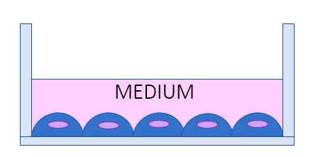
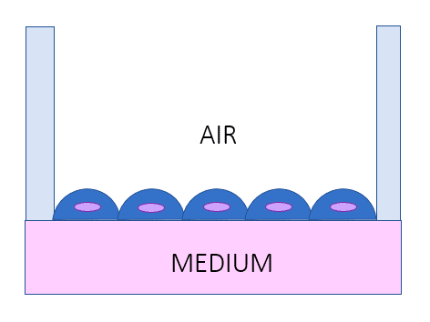
Organ-on-chip models vs. AIR-LIQUID INTERFACE LUNG MODEL [1, 5]

cell-organ-on-chip-in vitro-in vivo-drug testing-Microfluidics-Elveflow-Startup-Innovation-Technology

 +

  • Biomimetic stimuli and the possibility of cocultures
  • More representative phenotypes compared to 2D models

 –

  • Lack of 3D environment
  • Not dynamic environment

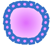
Organ-on-chip models vs. ORGANOIDS [6, 7]

organoid-organ-on-chip-in vitro-in vivo-drug testing-Microfluidics-Elveflow-Startup-Innovation-Technology

 +

  • 3D microenvironment
  • Used to study stem cell behavior
  • Different cellular types are included
  • Can mimic organ development, early-stage disease mechanisms, and tumorigenesis

 –

  • Lack of immune and vascular components
  • Lack of control of stem cells’ behavior and few reproducibility

Organ-on-chip models vs. TISSUE ENGINEERING (1)

tissue culture-organ-on-chip-in vitro-in vivo-drug testing-Microfluidics-Elveflow-Startup-Innovation-Technology

Artificial constructs [8-10]

 +

  • Reproducibility and a wide range of fabrication techniques
  • Several materials can be used (natural/ synthetic polymers, nanofibers, …)

 –

  • Difficult to mimic structural/ mechanical forces
  • Non autonomous production of ECM components

Organ-on-chip models vs. TISSUE ENGINEERING (2)

lung-organ-on-chip-in vitro-in vivo-drug testing-Microfluidics-Elveflow-Startup-Innovation-Technology

Biological (decellularized) constructs [8–10]

 +

  • Ability to model ECM-cell interactions
  • Complex 3D architecture is preserved
  • Presence of vascular structure

 –

  • Difficult to seed cells and reconstruct the functionality
  • The decellularization method can affect/ damage the device
  • Variability among species

Organ-on-chip models as solutions for the limitations of traditional in vitro and in vivo systems

Advantages resulting from the fabrication process

  • Modularity and variety of techniques/ materials for microfabrication
  • The chip design can be easily created and modified based on need
  • High reproducibility of fabrication protocols
  • Possibility to integrate sensors for high-throughput analyses
  • Possibility to develop personalized models

Advantages resulting from a microscale flow

  • Mimic physiological/diseased mechanical cues at a cellular/ tissue level
  • Create a controlled and dynamic microenvironment
  • Recreate the air-liquid interface (specifically for lungs)

Advantages for the cellular/ biological component

  • Create a complex tissue-like environment with several cell types
  • Create modular devices mimicking different organ areas (ex, bronchi, alveoli, etc.…in the case of lungs)
  • Reproducing physiological and pathological conditions

Advantages for time/cost of research

  • Fast fabrication processes (soft lithography, rapid prototyping, …)
  • Possibility to rapidly test drugs/compounds and have high-throughput outcomes.
  • Possibility to easily mimic disease conditions without the need to prepare, maintain, and use animal models.
eu_funded_en

Review done thanks to the support of the DELIVER H2020-MSCA-ITN-2017-Action “Innovative Training Networks.”

Grant agreement number: 766181

Written by Alessandra Dellaquila, PhD

Contact: 

Partnership@microfluidic.fr

References
  1. A. J. Miller and J. R. Spence, “In vitro models to study human lung development, disease and homeostasis,” Physiology, vol. 32, no. 3, pp. 246–260, 2017.
  2. B. A. Hassell et al., “Human organ chip models recapitulate orthotopic lung cancer growth, therapeutic responses, and tumor dormancy in vitro,” Cell Rep., vol. 21, no. 2, pp. 508–516, 2017.
  3. C. Blume and D. E. Davies, “In vitro and ex vivo models of human asthma,” Eur. J. Pharm. Biopharm. Off. J. Arbeitsgemeinschaft Pharm. Verfahrenstechnik EV, vol. 84, no. 2, pp. 394–400, Jun. 2013.
  4. H. Behrsing et al., “Assessment of in vitro COPD models for tobacco regulatory science: Workshop proceedings, conclusions and paths forward for in vitro model use,” Altern Lab Anim, vol. 44, no. 2, pp. 129–166, 2016.
  5. R. Bhowmick and H. Gappa-Fahlenkamp, “Cells and Culture Systems Used to Model the Small Airway Epithelium,” Lung, vol. 194, no. 3, pp. 419–428, Jun. 2016.
  6. K. Gkatzis, S. Taghizadeh, D. Huh, D. Y. R. Stainier, and S. Bellusci, “Use of three-dimensional organoids and lung-on-a-chip methods to study lung development, regeneration and disease,” Eur. Respir. J., vol. 52, no. 5, Nov. 2018.
  7. M. Huch, J. A. Knoblich, M. P. Lutolf, and A. Martinez-Arias, “The hope and the hype of organoid research,” Development, vol. 144, no. 6, pp. 938–941, Mar. 2017.
  8. A. Doryab, G. Amoabediny, and A. Salehi-Najafabadi, “Advances in pulmonary therapy and drug development: Lung tissue engineering to lung-on-a-chip,” Biotechnol. Adv., vol. 34, no. 5, pp. 588–596.
  9. R. Langer and J. Vacanti, “Advances in tissue engineering,” J. Pediatr. Surg., vol. 51, no. 1, pp. 8–12, Jan. 2016.
  10. D. M. Hoganson, E. K. Bassett, and J. P. Vacanti, “Lung tissue engineering,” Front. Biosci. Landmark Ed., vol. 19, pp. 1227–1239, Jun. 2014.

Stay in touch