Microfluidic reviews
|
Review done thanks to the support of the DeLIVER H2020-MSCA-ITN-2017-Action”Innovative Training Networks” – Grant agreement number: 766181 Author: Alessandra Dellaquila*, PhD candidate *corresponding author: Elvesys SAS, 172 Rue de Charonne 75011 Paris |
 |
1. Introduction on optical detection systems for microfluidics
 Suitable detection techniques are required to be coupled to microfluidic technology in order to analyze experiment outcomes in a sensitive and scalable way. The most common methods are optical detection techniques, electrochemical detection, mechanical analysis, spectroscopy methods (Raman spectroscopy, NMR spectroscopy) and mass spectrometry (MS) [1][2][3]. This review represents a brief overview of the imaging-based detection techniques and systems for microfluidics and a final mention of the latest and most innovative methods.
2. Optical detection systems for microfluidics
Several classifications of optical detection techniques can be done based on the optical method, the imaging systems or the presence or not of lenses [3][4]. Briefly, detection systems can be divided into off-chip (or free space systems)[5], in which detection components (light source, mirror and detector) are not integrated within the microfluidic device and light propagates in air, and integrated (on-chip) devices [6][7]. Optofluidic technology originates from integration of micro-optics and microfluidics [8]. Lenseless imaging can be performed through lens-free techniques [9][10]. Furthermore, emerging imaging techniques use nanoparticle labels and nanoengineered materials for optical detection [4].
Conventional and off-chip imaging methods
The use of bulk systems such as microscopes and interferometers is still widespread, thus microfluidics has been widely coupled to bright-field, phase-contrast, confocal and DIC microscopy [2][5] as well as interferometers [11]. Besides optical detection, also chemical imaging can be performed, usually by ATR-FTIR spectroscopy methods [12]. Optical detection is the dominant detection technique due to instrumentations availability and the ease to couple the systems to microfluidic platforms [13], however this devices are difficult to miniaturize and show reduced sensitivity due to short optical path lengths [4]. Also because of optics rapid development, optical detection provides nowadays for use of LEDs, lasers, diodes as light sources, microlenses, waveguides and optical fibers for detection and PMTs, CCDs, and CMOS as sensors [7].
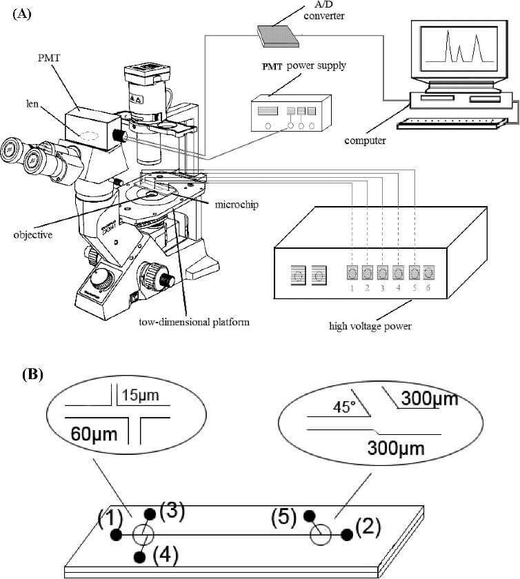
Figure 2. Use of inverted microscope and PMT detector to perform microchip electrophoresis in single red blood cells analysis[20].
Integrated imaging methods (on-chip imaging)
In on-chip imaging systems, optical and optoelectronic components are fully integrated within the microfluidic platform [1]. The main advantages of these systems in comparison with off-chip devices are operator-independence [6], increased portability, sensitivity, integration and the ability to tune the optical properties [14]. Furthermore, on-chip imaging can be performed through lens-free techniques such as shadow imaging or digital inline holography [9][15].
3. Optical detection techniques for microfluidic applications
The following table reports the most common imaging techniques. Based on the analytes, these techniques can be used for detection in analytical biochemistry (detection of proteins, cytometry, enzyme kinetics), POC diagnostics, cell biology, immunoassays as well as in screening applications and fluids manipulation[2][13][16]. Their application is not limited to the healthcare field but it is also extended to industrial and environmental areas.
| Technique | Principle | Properties |
| Absorbance | Measure of the attenuation of incident radiation in function of the wavelength | Simple instrumentation Low sensitivity |
| Fluorescence | Measure of emission light from a fluorophore | High sensitivity High selectivity Ease of incorporation |
| LIF and LEDIF | Excitation is caused by a laser or LED source | Focus on small volumes and low reagents amounts Single molecule detection |
| Chemiluminescence | Measure of energy release in a chemical reaction | High sensitivity and portability Does not require a light source Limited number of reagents Poor reproducibility |
| SPR | Measure of refractive index change of a sample in contact with a metal film | High sensitivity Complex and expensive instrumentation |
| Interferometers-based techniques | Measure of the phase shift caused by the analyte binding | High sensitivity Label-free |
| SERS | Measure of plasmonic effect of metal substrates or nanoparticles | High reliability and reproducibility High sensitivity |
Table 1. List of the most diffused optical techniques, their operating principle and characteristics [4][6] [7].
[starter_pack_rebound]
4. Optical detection components for microfluidics
Light sources
Commercial LEDs and miniaturized diode lasers as light sources have been widely used in microfluidics setup since their availability and low cost while organic LEDs (OLEDs) and dye lasers are the most widely used optofluidic light sources in current literature because the ease to be fully integrated within microfluidic systems and their versatility [13].
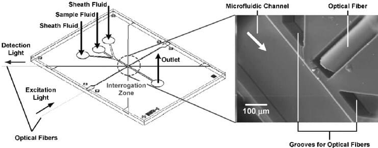
Figure 3. Use of embedded optical fibers for fluorescent measurements in a proteins immunoassay [13].
Optical components to increase detection
A standard and diffused approach is to use optical fibers, that can both transmit and detect light, are easy to come by and suitable for combination with other optical components. Absorbance, fluorescence and interferometry measurements can be improved by introduction of microlenses and waveguides. Waveguides can be classified as evanescent wave-based, such as liquid-core waveguides (LCWs) or interference based, as in the case of photonic crystals ARROWs detectors [6].
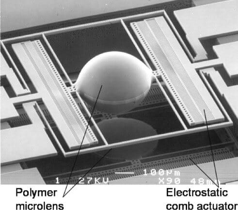
Figure 4. Polymer microlens for focusing and scanning [21].
Detectors
Optical detection can be performed by using a wide range of different detectors; the conventional ones are PMTs, CCD sensors, where CCDs allow also to perform multiplexing [4]. Integrated detectors are usually photodiodes (silicon or organic-OPDs) and CMOS sensors, that also permit lens-free imaging.
5. New perspectives for optical detection systems in microfluidics
As the increasing demand for cheap, automated, robust and portable platforms for a wide range of applications [17], sensitive and scalable optical detectors are required. As described by Myers et al., a new class of imaging detectors can be identified in nano-engineered probes, such as quantum dots (QDs), nanoparticles and biosensors [4]. Interestingly, development of smartphone-based microfluidic platforms enables to move towards fast, low-cost and simple analysis by using the smartphone camera as both light source and detector [18]. Moreover, efforts have been made to develop platforms with imaging systems based on high-speed optical technology [19] and nanoscopy to go beyond the spatial and temporal limits of conventional optical detection methods.
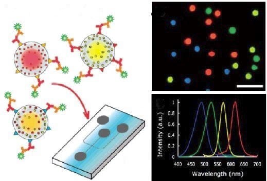
Figure 5. Schematic fluorescent image and emission spectra of QDs for immunosensing application [4].
Abbreviations: ARROW, antiresonant reflecting optical waveguides; CCD, charge-coupled device, CMOS, complementary metal-oxide-semiconductor; LCW, liquid-core waveguide; LED, light-emitting diode; LEDIF, LED-induce fluorescence LIF, laser-induced fluorescence; OPD, organic photodiode; PC, photonic crystal; PMT photomultiplier tube; POC, point-of-care; SERS, surface enhanced Raman spectroscopy; SPR surface plasmon resonance.
References
- K. B. Mogensen, H. Klank, and J. P. Kutter, “Recent developments in detection for microfluidic systems,” Electrophoresis, vol. 25, no. 21–22, pp. 3498–3512, 2004.
- Y. Zhu and Q. Fang, “Analytical detection techniques for droplet microfluidics-A review,” Anal. Chim. Acta, vol. 787, pp. 24–35, 2013.
- J. Wu and M. Gu, “Microfluidic sensing: state of the art fabrication and detection techniques,” J. Biomed. Opt., vol. 16, no. 8, p. 80901, 2011.
- F. B. Myers and L. P. Lee, “Innovations in optical microfluidic technologies for point-of-care diagnostics,” Lab Chip, vol. 8, no. 12, p. 2015, 2008.
- J. Wu, G. Zheng, and L. M. Lee, “Optical imaging techniques in microfluidics and their applications,” Lab Chip, vol. 12, no. 19, p. 3566, 2012.
- H. Gai, Y. Li, and E. S. Yeung, “Optical detection systems on microfluidic chips,” in Microfluidics, Springer, 2011, pp. 171–201.
- B. Kuswandi, Nuriman, J. Huskens, and W. Verboom, “Optical sensing systems for microfluidic devices: A review,” Anal. Chim. Acta, vol. 601, no. 2, pp. 141–155, 2007.
- X. Fan and I. M. White, “Optofluidic microsystems for chemical and biological analysis,” Nat. Photonics, vol. 5, no. 10, pp. 591–597, 2011.
- Z. Gorocs and A. Ozcan, “On-chip biomedical imaging,” IEEE Rev. Biomed. Eng., vol. 6, pp. 29–46, 2013.
- S. J. Moon, H. O. Keles, Y. G. Kim, D. Kuritzkes, and U. Demirci, “Lensless imaging for point-of-care testing,” Proc. 31st Annu. Int. Conf. IEEE Eng. Med. Biol. Soc. Eng. Futur. Biomed. EMBC 2009, vol. 24, pp. 6376–6379, 2009.
- Y.-C. Ahn, W. Jung, and Z. Chen, “Optical sectioning for microfluidics: secondary flow and mixing in a meandering microchannel,” Lab Chip, vol. 8, no. 1, pp. 125–133, 2008.
- K. L. A. Chan, S. Gulati, J. B. Edel, A. J. de Mello, and S. G. Kazarian, “Chemical imaging of microfluidic flows using ATR-FTIR spectroscopy,” Lab Chip, vol. 9, no. 20, p. 2909, 2009.
- H. Yang and M. A. M. Gijs, “Micro-optics for microfluidic analytical applications,” Chem. Soc. Rev., vol. 47, pp. 1391–1458, 2018.
- D. Psaltis, S. R. Quake, and C. Yang, “Developing optofluidic technology through the fusion of microfluidics and optics,” Nature, vol. 442, no. 7101, p. 381, 2006.
- M. Roy, G. Jin, D. Seo, M. H. Nam, and S. Seo, “A simple and low-cost device performing blood cell counting based on lens-free shadow imaging technique,” Sensors Actuators, B Chem., vol. 201, pp. 321–328, 2014.
- N. M. M. Pires, T. Dong, U. Hanke, and N. Hoivik, “Recent developments in optical detection technologies in lab-on-a-chip devices for biosensing applications,” Sensors (Switzerland), vol. 14, no. 8, pp. 15458–15479, 2014.
- P. Abgrall and A. M. Gué, “Lab-on-chip technologies: Making a microfluidic network and coupling it into a complete microsystem – A review,” J. Micromechanics Microengineering, vol. 17, no. 5, 2007.
- S. C. Kim, U. M. Jalal, S. B. Im, S. Ko, and J. S. Shim, “A smartphone-based optical platform for colorimetric analysis of microfluidic device,” Sensors Actuators, B Chem., vol. 239, pp. 52–59, 2017.
- K. Goda et al., “High-throughput single-microparticle imaging flow analyzer,” Proc. Natl. Acad. Sci., vol. 109, no. 29, pp. 11630–11635, 2012.
- S. Zhao, X. Li, and Y.-M. Liu, “Integrated Microfluidic System with Chemiluminescence Detection for Single Cell Analysis after Intracellular Labeling,” Anal. Chem., vol. 81, no. 10, pp. 3873–3878, 2009.
- S. Kwon and L. P. Lee, “Micromachined transmissive scanning confocal microscope,” Opt. Lett., vol. 29, no. 7, pp. 706–708, 2004.
For more information or Technical discussion

Microfluidics knowledge
Do you want tips on how to best set up your microfluidic experiment? Do you need inspiration or a different angle to take on your specific problem? Well, we probably have an application note just for you, feel free to check them out!


 Job
Job Collaborations
 Customer
 Other
Other